
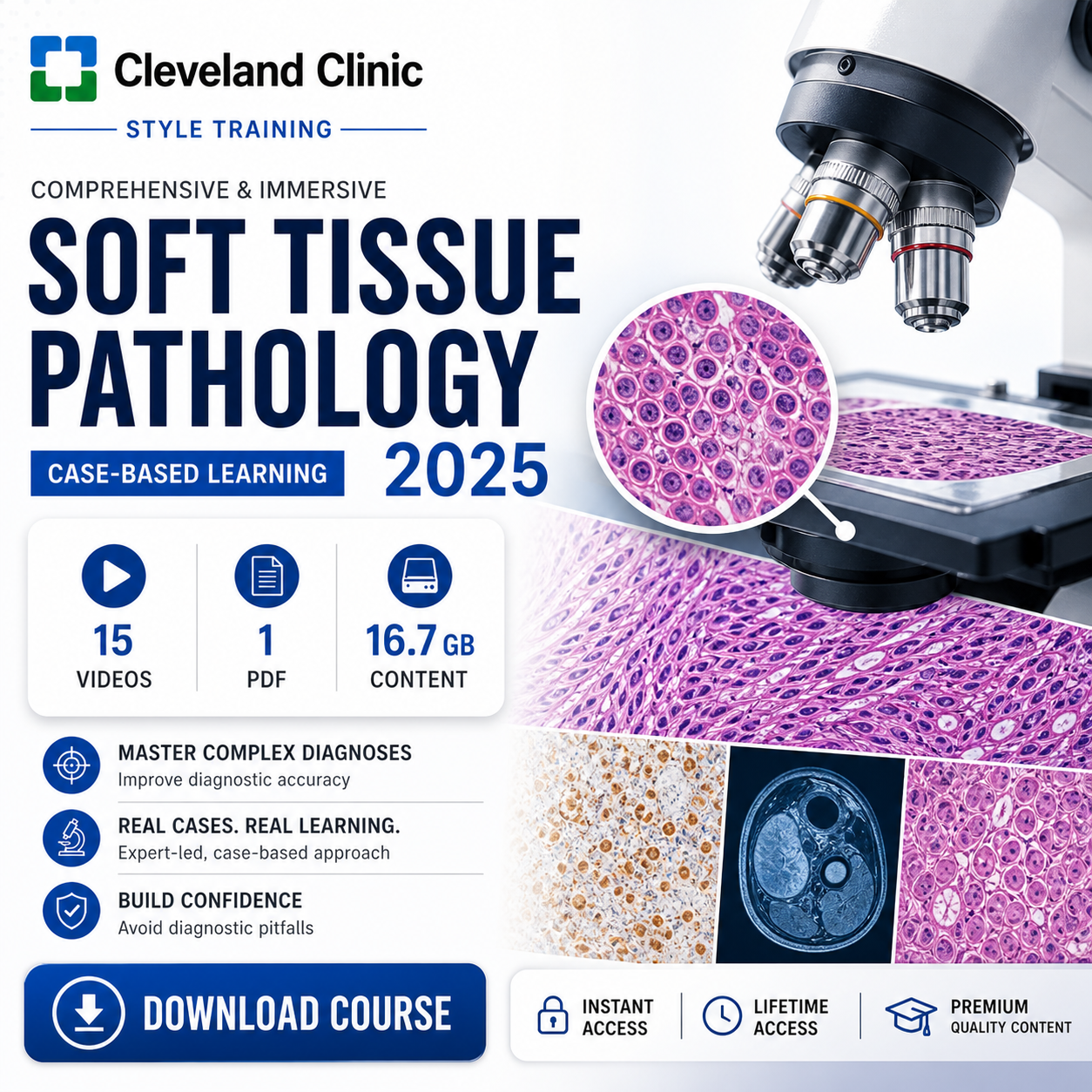
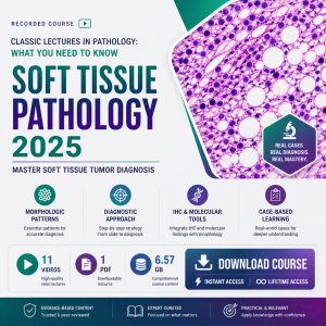

Cleveland Clinic Comprehensive and Immersive Soft Tissue Pathology 2025
A comprehensive case-based soft tissue pathology course focused on the practical diagnosis and work-up of mesenchymal lesions, from benign soft tissue tumors and reactive mimics to high-grade sarcomas, with emphasis on immunohistochemistry, molecular testing, radiology correlation, core biopsy scenarios, diagnostic pitfalls, and real-world pathology decision-making.
Course Details:
Cleveland Clinic Comprehensive and Immersive Soft Tissue Pathology 2025 includes 15 recorded videos and 1 PDF resource, offering an in-depth, case-based exploration of soft tissue pathology for pathologists, pathology residents, and fellows.
Focus: Lipomatous lesions, MDM2 FISH, lipomatous tumors, spindle cell sarcomas, herringbone and fascicular morphology, pseudosarcomas, acral soft tissue tumors, core biopsy scenarios, pediatric soft tissue pathology, periprosthetic joint infection, myxoid soft tissue tumors, S100-positive mesenchymal neoplasms, cutaneous soft tissue tumors, molecular correlation, mesenchymal tumors of the thorax and lung, and myoepithelioma differential diagnosis.
About This Course
Soft tissue pathology can be diagnostically challenging because mesenchymal lesions often show overlapping morphology, broad differential diagnoses, rare entities, reactive mimics, and important ancillary testing considerations.
Cleveland Clinic Comprehensive and Immersive Soft Tissue Pathology 2025 provides a practical, case-based approach to diagnosing soft tissue tumors. The course previews a wide range of real-world cases and highlights strategies for navigating common diagnostic challenges in daily pathology practice.
The program is designed to strengthen the pathologist’s ability to triage mesenchymal lesions, develop systematic work-up strategies, integrate immunohistochemistry and molecular tools, and correlate pathology findings with radiologic and clinical information.
Why Choose This Course
- Cleveland Clinic 2025 comprehensive and immersive soft tissue pathology course
- Includes 15 recorded videos and 1 PDF resource
- Case-based exploration from benign soft tissue lesions and reactive mimics to high-grade sarcomas
- Focuses on practical diagnostic strategies for mesenchymal lesions
- Strengthens use of immunohistochemistry, molecular techniques, and radiologic correlation
- Includes common and rare soft tissue tumors, diagnostic pitfalls, and systematic work-up approaches
- Useful for pathologists, pathology residents, fellows, and clinicians involved in soft tissue tumor diagnosis
Core Learning Areas
Mesenchymal Tumor Work-Up
Systematic diagnostic approach to mesenchymal neoplasms, biopsy triage, morphology patterns, ancillary test selection, and diagnostic workflow.
IHC & Molecular Tools
Immunohistochemistry, MDM2 FISH, molecular correlation, S100-positive neoplasms, molecular testing strategy, and integration of ancillary results.
Common & Rare Tumors
Lipomatous tumors, spindle cell sarcomas, myxoid soft tissue tumors, acral tumors, pediatric tumors, cutaneous tumors, and thoracic mesenchymal tumors.
Pitfalls & Mimics
Pseudosarcomas, reactive mimics, core biopsy limitations, diagnostic traps, infection-related pathology, and challenging differential diagnosis.
Complete Curriculum
- Case-based approach to soft tissue pathology
- Benign lesions, reactive mimics, and high-grade sarcomas
- Development of a systematic diagnostic approach
- Recognition of common and rare soft tissue tumors
- Practical work-up of mesenchymal lesions
- Integration of morphology, IHC, molecular testing, and radiology
- Recognition of diagnostic pitfalls in real-world pathology practice
- The utility of MDM2 FISH in the work-up of lipomatous lesions
- When MDM2 testing is helpful in differential diagnosis
- Distinguishing benign lipomatous lesions from atypical lipomatous tumor and well-differentiated liposarcoma
- Practical interpretation of ancillary testing results
- Common diagnostic pitfalls in lipomatous tumor work-up
- Correlation of morphology with molecular findings
- Case-based application of lipomatous lesion testing strategies
- Diagnostic approach to lipomatous tumors
- Recognition of common lipomatous neoplasms
- Atypical lipomatous tumor and liposarcoma considerations
- Morphologic patterns in adipocytic lesions
- When to use immunohistochemistry and molecular testing
- Distinguishing benign from malignant adipocytic tumors
- Practical reporting and diagnostic communication in lipomatous lesions
- Spindle cell sarcoma diagnostic framework
- Herringbone morphology and fascicular growth patterns
- Differential diagnosis of spindle cell mesenchymal tumors
- Use of morphology to guide ancillary testing
- Recognizing aggressive features and high-grade sarcoma patterns
- Immunohistochemical and molecular work-up considerations
- Case-based approach to spindle cell sarcoma diagnosis
- Recognition of pseudosarcomatous lesions
- Reactive mimics of sarcoma
- Clinical and histologic clues that prevent overdiagnosis
- Distinguishing benign reactive processes from malignancy
- Common diagnostic traps in soft tissue pathology
- Ancillary testing in challenging pseudosarcoma cases
- Practical strategies for avoiding misclassification
- Acral soft tissue tumor patterns
- Diagnostic clues in hand, foot, and distal extremity lesions
- Common acral tumor entities and mimics
- Clinical-pathologic correlation in acral lesions
- Case-based approach to acral tumor diagnosis
- Ancillary testing in diagnostically difficult acral tumors
- Practical pearls for recognizing rare acral soft tissue tumors
- Core biopsy triage in soft tissue pathology
- Limitations of small biopsy interpretation
- Prioritizing differential diagnosis on limited tissue
- Selecting the most useful ancillary tests
- Communicating uncertainty and recommending additional sampling
- Integrating radiology and clinical information with core biopsy findings
- Practical diagnostic strategies for common core biopsy scenarios
- Soft tissue tumors in pediatric patients
- Age-specific diagnostic considerations
- Benign and malignant pediatric mesenchymal lesions
- Recognition of pediatric tumor patterns
- Molecular and genetic considerations in pediatric tumors
- Differentiating developmental, reactive, and neoplastic processes
- Practical approach to pediatric soft tissue tumor work-up
- Pathologic evaluation of periprosthetic joint infection
- Histologic features supporting infection diagnosis
- Inflammatory patterns in periprosthetic tissue
- Correlation with clinical, microbiologic, and operative findings
- Diagnostic challenges in arthroplasty-related pathology
- Practical reporting considerations
- Role of pathology in multidisciplinary periprosthetic joint infection management
- Approach to myxoid soft tissue tumor differential diagnosis
- Recognition of benign and malignant myxoid lesions
- Morphologic patterns in myxoid mesenchymal tumors
- Immunohistochemical work-up of myxoid tumors
- Molecular testing strategies in myxoid neoplasms
- Common pitfalls in myxoid tumor diagnosis
- Case-based review of practical diagnostic decision-making
- Differential diagnosis of S100-positive mesenchymal tumors
- Peripheral nerve sheath tumors and related mimics
- Use of immunohistochemistry in S100-positive lesions
- Distinguishing neural, melanocytic, myoepithelial, and other tumor categories
- Ancillary markers that refine diagnosis
- Common pitfalls in S100 interpretation
- Systematic approach to S100-positive soft tissue neoplasms
- Recent updates in soft tissue tumor classification
- New entities and evolving diagnostic categories
- Emerging molecular findings in mesenchymal neoplasia
- Updated ancillary testing strategies
- Diagnostic implications of newly recognized tumor patterns
- Practical impact of updates on daily pathology practice
- Case-based discussion of current soft tissue pathology advances
- Common cutaneous soft tissue tumors encountered in practice
- Benign, atypical, and malignant cutaneous mesenchymal lesions
- Clinical-pathologic correlation in skin-based soft tissue tumors
- Distinguishing common tumors from important mimics
- Use of immunohistochemistry in cutaneous mesenchymal lesions
- Reporting considerations for everyday cutaneous soft tissue tumors
- Practical diagnostic pearls for dermatopathology and surgical pathology workflows
- Importance of molecular correlation in mesenchymal neoplasia
- Using molecular results to support or refine diagnosis
- When molecular testing is most useful in soft tissue tumors
- Integrating molecular findings with morphology and IHC
- Avoiding overreliance on isolated molecular results
- Communicating molecular-pathologic correlation in reports
- Practical framework for molecular testing in soft tissue pathology
- Mesenchymal tumors involving thorax and lung
- Clinical and radiologic correlation in thoracic soft tissue lesions
- Differential diagnosis of pulmonary and thoracic mesenchymal tumors
- Benign, intermediate, and malignant entities
- Immunohistochemical and molecular work-up strategies
- Diagnostic pitfalls in thoracic mesenchymal pathology
- Practical approach to thoracic and lung-based mesenchymal neoplasms
- Myoepithelioma diagnostic considerations
- Soft tissue myoepithelial tumor differential diagnosis
- Recognizing morphologic variability in myoepithelial lesions
- Immunohistochemical work-up of suspected myoepithelioma
- Molecular correlation in difficult myoepithelial tumors
- Common mimics and diagnostic traps
- Case-based strategy for deciding “myoepithelioma or not”
Learning Objectives
- Comprehend the utility of immunohistochemical, molecular, and radiology studies in the work-up of soft tissue tumors
- Develop a systematic work-up strategy for the diagnosis of mesenchymal neoplasms
- Identify common and uncommon soft tissue tumors across multiple anatomic sites
- Recognize diagnostic pitfalls commonly encountered in soft tissue pathology
- Use MDM2 FISH appropriately in the evaluation of lipomatous lesions
- Improve diagnostic confidence in spindle cell, myxoid, acral, pediatric, cutaneous, thoracic, and S100-positive mesenchymal tumors
- Distinguish true sarcomas from pseudosarcomas and reactive mimics
- Apply practical strategies for limited core biopsy scenarios in soft tissue pathology
- Integrate molecular correlation into diagnosis and reporting of mesenchymal neoplasms
- Translate case-based diagnostic pearls into everyday surgical pathology practice
Who Should Take This Course
This course is designed for pathologists, pathology residents, and fellows who want to strengthen their diagnostic approach to soft tissue pathology and mesenchymal neoplasms.
It is especially useful for learners who evaluate soft tissue tumors, sarcoma biopsies, cutaneous mesenchymal lesions, pediatric soft tissue lesions, thoracic mesenchymal tumors, molecular pathology results, IHC panels, and diagnostically challenging surgical pathology cases.
Delivery & Access
15 Videos
Recorded case-based lectures covering essential soft tissue pathology topics.
1 PDF
Supporting PDF material included for review and reference.
16.7 GB
Complete course package with recorded video learning materials.
Support
Course Highlights
Case-Based Diagnosis
Real-world soft tissue pathology cases covering benign lesions, reactive mimics, uncommon tumors, and high-grade sarcomas.
Ancillary Testing Focus
Strong emphasis on immunohistochemistry, MDM2 FISH, molecular correlation, radiologic correlation, and systematic diagnostic work-up.
Practical Pathology Pearls
Helps pathologists recognize pitfalls, triage core biopsies, and approach challenging mesenchymal neoplasms more confidently.
FAQ
Who is this course best suited for?
This course is best suited for pathologists, pathology residents, and fellows who want a practical, case-based review of soft tissue pathology.
What is included in this course?
The course includes 15 recorded videos and 1 PDF resource, with a complete package size of 16.7 GB.
What topics are covered?
Topics include MDM2 FISH in lipomatous lesions, lipomatous tumors, spindle cell sarcomas, pseudosarcomas, acral soft tissue tumors, core biopsy scenarios, pediatric soft tissue pathology, periprosthetic joint infection, myxoid soft tissue tumors, S100-positive mesenchymal neoplasms, soft tissue pathology updates, cutaneous soft tissue tumors, molecular correlation, thoracic and lung mesenchymal tumors, and myoepithelioma differential diagnosis.
Does this course cover immunohistochemistry and molecular testing?
Yes. The course emphasizes immunohistochemistry, molecular techniques, MDM2 FISH, molecular correlation, and integration of ancillary testing into soft tissue tumor diagnosis.
Does this course help with diagnostic pitfalls?
Yes. The course focuses on recognizing common pitfalls in soft tissue pathology, including reactive mimics, pseudosarcomas, small biopsy limitations, and challenging differential diagnoses.
Is this a recorded course?
Yes. This product is provided as recorded course access so you can review the material at your own pace.
Does this course include CME or a certificate?
No. CME credits and certificates are not included with this course package.
Who can I contact for support?
You can contact support at [email protected].
Reviews
There are no reviews yet.