
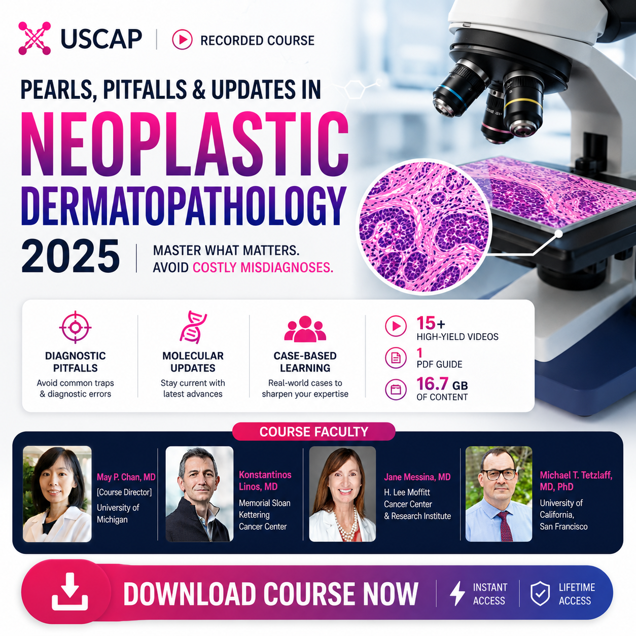



USCAP Pearls, Pitfalls, and Updates in Neoplastic Dermatopathology 2025
A focused USCAP dermatopathology course covering major areas in neoplastic dermatopathology, including melanocytic, mesenchymal, adnexal, epithelial, neuroendocrine, and hematolymphoid neoplasms of the skin, with a case-based approach to diagnostic challenges, pitfalls, molecular updates, and ancillary testing.
Course Details:
USCAP Pearls, Pitfalls, and Updates in Neoplastic Dermatopathology 2025 is a recorded case-based dermatopathology course designed for practicing academic pathologists, community pathologists, and pathologists-in-training.
Focus: Melanocytic neoplasms, adnexal neoplasms, mesenchymal neoplasms, epidermal neoplasms, neuroendocrine tumors, hematolymphoid neoplasms of the skin, newly described cutaneous tumors, molecular aberrations, histopathologic features, immunophenotypes, ancillary studies, diagnostic pitfalls, skin biopsy interpretation, and practical dermatopathology decision-making.
About This Course
Neoplastic dermatopathology is one of the most challenging areas of diagnostic pathology because many cutaneous tumors show overlapping histologic patterns, subtle immunophenotypic clues, molecularly defined entities, and important diagnostic pitfalls.
This USCAP 2025 course uses a case-based didactic approach to help learners recognize classic and subtle features that distinguish between different cutaneous neoplasms. The faculty focus on practical diagnostic pearls, common traps, new entities, and the appropriate use of ancillary studies.
The course is designed to strengthen diagnostic confidence in skin biopsy interpretation, especially in complex neoplastic dermatopathology cases involving melanocytic, adnexal, mesenchymal, epithelial, neuroendocrine, and hematolymphoid tumors.
Why Choose This Course
- USCAP 2025 course focused on pearls, pitfalls, and updates in neoplastic dermatopathology
- Case-based didactic approach for real-world dermatopathology diagnosis
- Covers melanocytic, mesenchymal, adnexal, epidermal, neuroendocrine, and hematolymphoid neoplasms of the skin
- Highlights classic and subtle histopathologic features used to distinguish cutaneous neoplasms
- Reviews immunophenotypes, ancillary studies, molecular aberrations, and newly described cutaneous tumors
- Emphasizes common diagnostic challenges, pitfalls, and practical ways to avoid misinterpretation
- Designed for practicing academic and community pathologists, dermatopathologists, pathology residents, and fellows
Core Learning Areas
Melanocytic Neoplasms
Diagnostic patterns, subtle histologic clues, immunophenotypes, molecular updates, difficult melanoma mimics, and practical pitfalls in melanocytic tumor interpretation.
Adnexal Neoplasms
Sweat gland, follicular, sebaceous, and adnexal tumor patterns with emphasis on accurate classification, differential diagnosis, and practical diagnostic clues.
Mesenchymal Neoplasms
Cutaneous mesenchymal tumors, spindle cell lesions, vascular tumors, soft tissue mimics, ancillary testing, and molecular correlation in skin-based neoplasms.
Epithelial & Hematolymphoid Tumors
Epidermal, neuroendocrine, and hematolymphoid neoplasms of the skin, including diagnostic pitfalls, immunostain strategy, and challenging biopsy scenarios.
Complete Curriculum
- Major areas in neoplastic dermatopathology
- Case-based didactic learning format
- Practical tips for accurate diagnosis
- Diagnostic challenges and pitfalls in skin biopsies
- Newly described cutaneous tumors
- Defining molecular aberrations in emerging entities
- Ancillary tools that aid diagnosis and help refine differential diagnosis
- Diagnostic approach to cutaneous adnexal tumors
- Recognition of follicular, sebaceous, apocrine, and eccrine tumor patterns
- Benign versus malignant adnexal neoplasm features
- Key histologic clues for adnexal tumor classification
- Immunohistochemical support in adnexal tumor diagnosis
- Common diagnostic pitfalls in adnexal neoplasms
- Case-based discussion of challenging adnexal tumors
- Classic and subtle histopathologic features of melanocytic neoplasms
- Differentiating benign, atypical, borderline, and malignant melanocytic lesions
- Melanoma mimics and diagnostic traps
- Immunophenotypic patterns in melanocytic tumor work-up
- Molecularly defined melanocytic entities
- Ancillary studies in difficult melanocytic lesions
- Practical pearls for improving melanoma diagnosis and avoiding overdiagnosis
- Epidermal neoplasms and important diagnostic patterns
- Cutaneous neuroendocrine neoplasms
- Hematolymphoid neoplasms involving the skin
- Immunophenotypic features that guide classification
- Distinguishing primary cutaneous tumors from secondary involvement
- Diagnostic pitfalls in epithelial, neuroendocrine, and hematolymphoid lesions
- Case-based approach to difficult skin biopsy interpretation
- Cutaneous mesenchymal tumor patterns
- Spindle cell lesions and soft tissue tumor mimics
- Vascular, fibrohistiocytic, neural, and other mesenchymal neoplasms
- Immunohistochemistry in mesenchymal tumor diagnosis
- Molecular correlation in skin-based mesenchymal tumors
- Distinguishing benign, intermediate, and malignant lesions
- Practical diagnostic approach to challenging mesenchymal skin biopsies
- Faculty discussion of complex neoplastic dermatopathology cases
- Diagnostic reasoning across multiple cutaneous tumor categories
- How experts approach challenging differential diagnoses
- When to use ancillary studies and when to recognize their limitations
- Common pitfalls in real-world dermatopathology practice
- Strategies for improving diagnostic accuracy in skin biopsy evaluation
- Practical takeaways for academic and community pathology workflows
- Classic morphologic features of common cutaneous neoplasms
- Subtle histologic findings that refine differential diagnosis
- Immunophenotypic patterns used to distinguish tumor categories
- Appropriate selection of immunohistochemical panels
- Interpreting immunostains in the context of morphology
- Recognizing misleading or nonspecific staining patterns
- Combining morphology and immunophenotype for accurate diagnosis
- New entities in cutaneous tumor pathology
- Histomorphologic features of recently described tumors
- Genetic aberrations that define emerging diagnostic categories
- Molecular findings that support tumor classification
- When molecular testing is useful in dermatopathology
- Integrating molecular data into diagnostic reports
- Practical implications of molecularly defined cutaneous neoplasms
- Appropriate use of immunohistochemistry
- Understanding the limitations of ancillary studies
- Ancillary tools in melanocytic tumor diagnosis
- Ancillary tools in adnexal tumor work-up
- Ancillary tools in mesenchymal neoplasm classification
- Ancillary tools in epithelial, neuroendocrine, and hematolymphoid neoplasms
- Using ancillary studies to resolve, not create, diagnostic uncertainty
- Common pitfalls in cutaneous neoplasm interpretation
- Overdiagnosis and underdiagnosis in dermatopathology
- Small biopsy limitations and sampling issues
- Misleading histologic patterns and overlapping features
- When clinical correlation is essential
- How to avoid misclassification of difficult skin tumors
- Practical strategies for improving diagnostic confidence and consistency
Learning Objectives
- Identify classic and subtle histopathologic features that distinguish between different cutaneous neoplasms
- Recognize immunophenotypic patterns that help classify neoplastic skin lesions
- Become familiar with newly described cutaneous tumors defined by unique histomorphologic features
- Understand genetic aberrations that define or support diagnosis of emerging cutaneous neoplasms
- Use ancillary studies appropriately in neoplastic dermatopathology
- Recognize the limitations of immunohistochemistry, molecular testing, and other ancillary tools
- Improve diagnostic accuracy in melanocytic, adnexal, mesenchymal, epithelial, neuroendocrine, and hematolymphoid skin tumors
- Recognize potential diagnostic pitfalls when evaluating skin biopsies
- Apply practical strategies to avoid overdiagnosis, underdiagnosis, and misclassification
- Translate case-based dermatopathology pearls into everyday academic and community pathology practice
Who Should Take This Course
This course is designed for practicing academic pathologists, community pathologists, dermatopathologists, pathology residents, fellows, and pathologists-in-training who evaluate neoplastic skin biopsies and cutaneous tumors.
It is especially useful for learners who want a practical, case-based review of melanocytic neoplasms, adnexal tumors, mesenchymal tumors, epithelial lesions, neuroendocrine tumors, hematolymphoid neoplasms, molecularly defined entities, ancillary testing, and diagnostic pitfalls in dermatopathology.
Delivery & Access
Recorded Course
Review the neoplastic dermatopathology sessions at your own pace.
2025 Edition
Updated dermatopathology content focused on current entities, pitfalls, and ancillary tools.
Case-Based Learning
Practical diagnostic teaching using real-world style dermatopathology cases.
Support
Course Highlights
Case-Based Diagnostic Focus
Uses practical cases to clarify complex neoplastic dermatopathology diagnosis and strengthen pattern recognition.
Modern Molecular Updates
Covers newly described cutaneous tumors, molecular aberrations, and ancillary tools that support accurate diagnosis.
Pitfalls & Pearls
Emphasizes common diagnostic traps and practical ways to avoid errors in skin biopsy interpretation.
FAQ
Who is this course best suited for?
This course is best suited for practicing academic pathologists, community pathologists, dermatopathologists, pathology residents, fellows, and pathologists-in-training.
What topics are covered?
Topics include melanocytic neoplasms, adnexal neoplasms, mesenchymal neoplasms, epidermal neoplasms, neuroendocrine tumors, hematolymphoid neoplasms of the skin, newly described cutaneous tumors, molecular aberrations, histopathologic features, immunophenotypes, ancillary studies, diagnostic pitfalls, and case-based skin biopsy interpretation.
Does this course cover melanocytic tumors?
Yes. The course includes melanocytic neoplasms, difficult melanocytic lesion diagnosis, immunophenotypic patterns, melanoma mimics, molecular updates, and practical diagnostic pitfalls.
Does this course cover ancillary testing?
Yes. The course emphasizes appropriate use and limitations of ancillary studies, including immunohistochemistry and molecular tools used in neoplastic dermatopathology.
Is this course case-based?
Yes. The course uses a case-based didactic approach to help learners recognize diagnostic clues, avoid pitfalls, and improve interpretation of neoplastic skin biopsies.
Is this a recorded course?
Yes. This product is provided as recorded course access so you can review the material at your own pace.
Does this course include CME or a certificate?
No. CME credits and certificates are not included with this course package.
Who can I contact for support?
You can contact support at [email protected].


Dr. Sarah Thompson: –
“This course provided invaluable insights that transformed my approach to complex cases!”
Dr. James Lee: –
“Expert presentations made challenging topics accessible and engaging.”
Dr. Maria Gonzalez –
“The case discussions were particularly helpful in clarifying my diagnostic uncertainties.”
Dr. Alan Chen –
“I appreciated the flexibility of recorded sessions on Google Drive—perfect for my schedule!”
Dr. Emily Patel –
“This course has significantly boosted my confidence in dermatopathology.”
Dr. Laura Hall –
“I feel much better equipped to handle complex dermatopathology cases after this course!”
Dr. Natalie Turner –
“I learned so much from the case studies; they were very enlightening.”
Dr. Brian Carter –
“This course has become a crucial resource for my continuing education.”
Dr. Rachel Adams –
“The course exceeded my expectations—fantastic content and delivery!”
Dr. Kevin Wilson: –
“I gained new perspectives on diagnosing neoplastic conditions that I hadn’t considered before.”
Dr. Daniel Nguyen –
“I highly recommend this course to anyone in the field of dermatopathology.”
Dr. Angela Martinez –
“Every session was packed with useful information that I could apply immediately.”
Dr. Chris Johnson –
“The interactive format kept me engaged throughout the course.”